الصفحة الرئيسية / المجموعات / الصفحة الرئيسية / Hospital Clinic Health Care Center 4D Color Doppler Ultrasound Machine DW-KT6 | DAWEI

Hospital Clinic Health Care Center 4D Color Doppler Ultrasound Machine DW-KT6 | DAWEI

$9,989.00
| /

Hospital Clinic Health Care Center 4D Color Doppler Ultrasound Machine DW-KT6 | DAWEI

$9,989.00
| /
وصف

Product name: Brand new CE Approved Trolley 4D Color Doppler Ultrasound Diagnostic System

High-precision ultrasound system designed specifically for women’s health


CE Certified Trolley 4D Baby Color Doppler Ultrasound Scan Machine DW-KT6:
☑️A full series of probes.
☑️Abundant obstetrics and gynecology measurement kit.
☑️Vascular applications: A variety of high frequency probes for vascular examination, supporting color and PW, tri-sync, acoustic beam deflection, Angle correction, 2-d/color synchronous display, and convenient measurement and reporting.
☑️Beam fan-shaped expansion technology: high speed and high quality without splicing.
☑️Cross transmit and receive: Eliminates side effects.
☑️Full digital UWB: improves resolution and penetration.
☑️Built-in graphic management system, the diagnostic report can be edited, ultrasound diagnosis image embedded in the report, and directly printed;
☑️Hard disk dynamic and static image storage, real-time sharing;
☑️Image formats:≥4;
☑️Input/output interfaces: HDMI interface, video output/input interface, S-Video, parallel print interface, network interface, USB interface, DISC.
☑️19-inch square screen + 10.3-inch touch screen.
☑️Fully activated probe port: 4
☑️Built-in 128G solid-state drive.
☑️Image optimization, Multi-Beam, IMT, Trapezoidal imaging.With Intelligent PIP imaging mode.
☑️Windows 8 Embedded operation system.


Transducer Applications
Convex Array *Recommended Abdomen, OB/GYN, Urology
Transvaginal Endovaginal, OB/GYN
Trans-rectal Urology
Micro-Convex Array Pediatrics
Linear Array *Recommended Small Parts, Vascular, Musculoskeletal, Superficial
4D volume OB/GYN
Phased array Cardiology

1)On the left side of the fuselage, there is a DISC insert. Is it for CD or DVD ?

This is the CD-ROM drive, used to burn discs, and can also read the CD-ROM drive of the printer.

2)  Can I transfer images through the USB port ?

Absolutely it can transfer images through the USB port. Insert the usb flash drive into the USB port, you can transfer the pictures in the machine to the usb flash drive.
There are Delete and Share buttons, Select a picture and click the Share button, and there will be shared channels, including the inserted usb flash drive.

3) How will I conduct this machine?

✅Operation instruction video, various documents including parameters and brochures
✅Our after-sales engineers are responsible for each machine sold, and provide free training for customers who do not know how to use the machine.
✅The training is usually conducted online through WhatsApp or zoom.
✅Lifetime after-sales | We will also provide assistance if you have any questions during use.

4) A question about the ultrasound machine does it works without internet?


All of our ultrasound machines can work without the internet.
-DW-KT6 is a system embedded with Windows 8, it can also work without the internet.
-We recommend that this ultrasound machine be used without internet connection, because the ultrasound software of the machine will be killed by mistake because of the Internet connection.
-The ultrasound machine does not have WiFi function.
-The built-in Windows system can be used to drive installation, software upgrades, etc.

T6 Clinical image

DW-KT6 Technical Parameters

1

Summary of main specifications and system of Trolley 4D color Doppler ultrasound

1.1

Trolley type all digital color Doppler ultrasonic mainframe

1.2

Ultrasonic host operating system: Windows 8 operating system

1.3

Spectrum pulse Doppler

1.4

Direction energy Doppler

1.5

Real time three synchronization

1.6

Space composite imaging: the requirement is 3 level, visual adjustable.

1.7

Organized harmonic imaging technology

1.8

4B imaging mode

1.9

One key intelligent optimization

1.10

System languages: Chinese/English/ Spanish/ French/ Russian

1.11

Monitor: 19 inches, high definition LED.

1.12

10.4 inches touch LED

1.13

Physical clipboard: save the image on the left side of the screen, which can be directly saved or deleted.

1.14

The system has the function of on-the-spot upgrade

1.15

Presupposition: for different inspection of the viscera, preset the inspection conditions for the best image, reduce the adjustment of the operation, and the commonly used external adjustment and combination regulation.

1.16

Support real-time 3D imaging function

1.17

The probe interface is 4

2

Probes

2.1

Convex array probe: 2.5MHz/3.0MHz/3.5MHz/4.0MHz/H4.0MHz/H5.0MHz,  

2.2

Linear array probe: 6.0MHz/7.5MHz/9.0MHz/12.0MHz/H10.0MHz,  

2.3

Trans-vaginal probe: 4.5MHz/6.0MHz/7.0MHz/9.0MHz/H8.0MHz,  

2.4

Phased array probe: 2.5MHz/3.0MHz/3.5MHz/4.0MHz/H3.0MHz/H4.0MHz,  

2.5

Micro convex probe: 4.5MHz/6.0MHz/7.0MHz/9.0MHz/H8.0MHz  

2.6

4D Volume probe: 2.0MHz/3.0MHz/4.5MHz/6.0MHz/H5.0MHz, 

2.7

The above probe has harmonic frequency

2.8

It can be selected according to customer needs: convex array probe, linear array probe, rectal probe, micro convex probe, phased array probe, volume probe.

2.9

Under each probe, there is a selection of specialist and viscera mode and rapid entry detection.

3

Two-dimensional imaging mode

3.1

Gain: 0-100, step 1 visible adjustable

3.2

TGC:8 segment adjustable

3.3

Image optimization: visible and adjustable over 7 levels

3.4

Dynamic range: 20-280dB 20 level visual adjustable

3.5

False color: 12, visible and adjustable

3.6

Smooth treatment: 8, visible and adjustable

3.7

Edge enhancement: 8, visible and adjustable

3.8

Sound power: 5% to 100%, step 5%, visible and adjustable

3.9

Display depth: greater than 317mm, less than 20mm, cardiac probe 371mm

3.10

Maximum focus number: 6 focal points, which can be moved throughout the whole process.

3.11

Scan line density more than 256 visible tunable

3.12

Gray scale: 0-7 level visible visible and adjustable

3.13

Filtering, 5 kinds

3.14

Scanning range, 50%-100%

3.15

Frame correlation, 0-4 level, visible and adjustable

3.16

The screen has real-time display of voice power, probe frequency, dynamic range, pseudo color, gray scale and other 14 parameters can be adjusted

4

Color imaging mode

4.1

Color frequency: 8 frequency conversion, visible adjustable

4.2

Color deflection: equipped with

4.3

Color frame correlation 12 levels, visible and adjustable

4.4

Color map: 7, visible and adjustable

4.5

Color reversal: adjustable

4.6

B/C split screen synchronous display function: equipped with

4.7

Color baseline: 7, visible and adjustable

4.8

Color line density: adjustable

5

Spectrum Doppler mode

5.1

Sampling volume angle correction: -80 degree to 80 degree adjustable

5.2

Sample volume: 0.5mm-20mm visually adjustable

5.3

Frequency: ≥5, visible and adjustable

5.4

Baseline: 8 adjustable

5.5

Smooth: 8 files can be adjusted

5.6

Display layout: 4 visible tunable

5.7

False color: 7 kinds of adjustable

5.8

Speed scale: 3-2288cm/s visibility adjustable

5.9

Spectrum envelope function: real time automatic spectrum envelope, manual spectrum envelope, and other modes. The system automatically analyses and displays various data such as PSV, EDV, RI, PI, S/D, ACC, HR and so on.

6

Measurement and analysis function:

6.1

General measurement distance, area, angle, time, slope, heart rate, velocity, acceleration, neck hyaline layer, spectrum tracing, resistance index / pulsatility index, etc.

6.2

Obstetric measurement: weight measurement formula more than 8 options

6.3

The color and line type of the measuring line can be adjusted at will (including activating the color and completing the color).

6.4

The measurement results show that the location and font size can be adjusted according to needs.

6.5

Professional software package: abdomen, volume, ratio, obstetrics and Gynecology, small organs, carotid artery, Urology, orthopedics, peripheral blood vessels, heart.

7

Graphic and text management system

7.1

Host built in ≥128G solid state hard disk to start fast and stable

7.2

Movie playback: 1200 frames

7.3

Internal file information management system: can record patient number, name, check number, check date and so on, and can be searched and managed by numbering, checking number, name and so on.

7.4

The type of report is 16

7.5

One key fast report graphic and text management

8

Interface

8.1

USB interface: 4

8.2

DVI interface: 1

8.3

RJ-45 interface: 1

8.4

VIDEO interface: 1

8.5

Grounding wire interface: 1

8.6

S-VIDEO interface: 1

8.7

HDMI interface: 1

9

Technology, after-sales service and other requirements

9.1

After acceptance, the warranty is free for two years

9.2

Manufacturer has ISO13485 certification and EU CE certification.


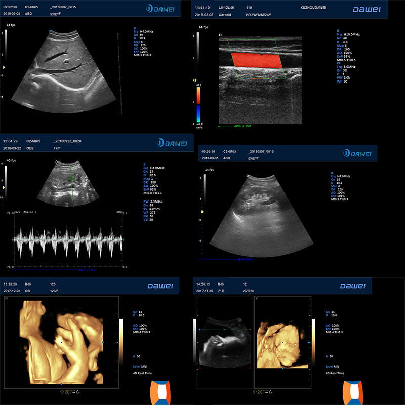
The full-body applied color Doppler ultrasound diagnosis system uses a new technology platform based on a safe and stable operating system to calmly face various emergencies. High performance, low power consumption industrial control solution, to ensure better image performance quality presentation. High brightness, high resolution color medical LIQUID crystal display. High sensitivity and large touch screen make doctors enjoy more operation fun in clinical application. Excellent imaging technology, ultrasound image accurate display, widely used in the abdomen, urology, obstetrics and gynecology, pediatrics/neonatology, superficial tissue, musculoskeletal, heart, etc. Extended imaging, not limited to this field of vision, better display effect, one-click access, deep customization of application types, checking habits without changing, update Windows platform design, richer functions, more stable performance.