

Good image quality is fairly subjective. It’s also relative to the capabilities of the machine and physicians operation experience. Getting started, everyone must know these basic controls. You’ll use these most often no matter how advanced you get.
- Don’t fear the gain control
The gain knob will likely be your most-used imaging control. This adjusts the overall brightness of the ultrasound image.
- TGC: Time Gain Compensation controls
With the exception of SonoSite and many tablets, each machine has 5-10 slide controls grouped together. These are TGC or Time Gain Compensation controls. They adjust gain in specific areas of the image The best way to see what a control does is to slide one of the controls all the way to the right, then all the way to the left while looking at a live image. You’ll see that a certain section of the image turns very bright, then very dark.
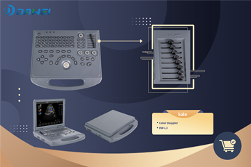
(click the picture for more details)
- Ultrasound Focus position, focal zones
These are two often-overlooked image optimization features. The focal position tells the ultrasound the depth at which you’d like the highest resolution. As you move the focal position up and down, you’ll see a triangle or dot move up or down the left or right side of the image. You’ll see the image resolution improve in the area of the selected focal position.
Focal zones allow you to have multiple focus points. As you increase the number of focal zones, your frame rate will decrease and the image will refresh slowly.
- Frequency Selection
Adjusting the frequency allows you to increase resolution at the expense of penetration, or increase penetration at the expense of resolution.
Use lower frequencies when you need penetration in the image. Use higher frequencies when you are looking at superficial imaging. High frequencies provide the best resolution, but you lose penetration. Low frequencies provide the best penetration at the expense of image resolution.
- Auto Optimization
Many ultrasounds come with a feature that automatically optimizes the gain and overall contrast of the image. This feature analyzes the tissue in the image and attempts to provide you with the most optimized image.
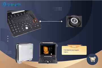
(click the picture for more details)
- Line Density
Line density adjusts the number of scan lines in your ultrasound image. A higher level provides better resolution in the image (more scan lines), but reduces the frame rate. Use this to get the best possible image with the most acceptable frame rate.
- Dynamic Range (Compression)
Dynamic Range (also known as Compression) allows you to tell the ultrasound machine how you want the echo intensity displayed as shades of gray. A broad/wide range will display more shades of gray and an overall smoother image. A smaller/narrow range will display fewer shades of gray and appear as a higher contrast with a more black-and-white image.
- Tissue Harmonics Imaging(THI)
This is the most common advanced imaging technology and is found on about 90% of available new and refurbished ultrasound machines.
Harmonics imaging allows the ultrasound to identify body tissue and reduce artifact in the image. It does this by sending and receiving signals at two different ultrasound frequencies.
- Speckle Reduction Imaging (SRI)
Speckle Reduction Imaging uses an algorithm to identify strong and weak ultrasound signals. By evaluating the image on a pixel-by-pixel basis, it attempts to identify tissue and eliminate “speckle.” Weak signals that seem to be astray are eliminated, while strong signals are enhanced/brightented. It provides a smoother, cleaner image.
Above are all we summarized. Try to use those function buttons to better improve the dignostic buttons. Besides, more advanced image improvement technique will be updated in the near future. Dawei Medical is always providing the advanced clinical imaging experience together with after-sale training and supports!