Colour-flow duplex imaging (CFDI) investigation has become the cornerstone of the diagnostic-therapeutic approach to varicose vein disease; morphologic and haemodynamic information may be safely collected with great accuracy by means of duplex (colour-flow) exploration, reducing greatly the necessity of invasive contrastographic means.
In the last few years it has been possible to highlight a few "new" patterns of saphenous incompetence: a) long and short saphenous vein are very rarely (3% of the affected limbs in cases of LSV) insufficient from the junction to the ankle, a segmental reflux being more common, b) in cases of long saphenous reflux, the sapheno-femoral junction is competent (no reflux from common femoral vein) in about 30% of the patients, while several extra-junctional reverse flows are possible, c) in primary varicose veins perforating veins of the leg have mainly a re-entry role, even if enlarged and with bi-directional flow.
In the field of superficial venous disease, CFDI permits also: a) the clarification of the vast majority of the complex patterns of recurrent varices, b) the definition and the monitoring of the superficial venous thrombosis, finally c) improvement in our knowledge of the minor varicosities and finally even d) the possibility to guide a safer and more precise sclerotherapic session (ultrasound guided sclerotherapy).
Whatever the therapeutic plan may be, the treatment of varicose vein disease must be guided by a correct diagnostics, principally relying upon colour-flow-duplex imaging: the true gold standard for this kind of vascular disease.

Ultrasound diagnostics, which began with doppler c.w. and then duplex scanning (DS), have into the last few years developed into colour-flow duplex imaging (CFDI), which guarantees better anatomical and functional accuracy.
Such safe, non invasive, and reliable equipment has progressively revolutionized all the varicose vein disease diagnostics and has greatly improved surgery and sclerotherapy (1). Nowadays DS or CFDI may (and must) guide the phlebologist's therapeutical steps, when treating trunk varicose veins or smaller varicosities of the lower limbs. By means of this kind of examination it is possible to collect several, precious anatomic information about the different characteristics of the superficial venous system (SVS), the deep venous system (DVS) and the perforating veins (PV) of the lower limb.
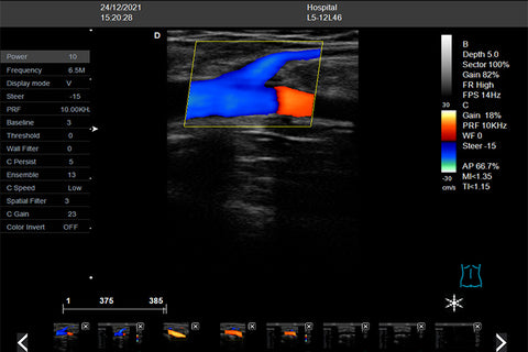
Alongside morphological information, it is possible to study the haemodynamics of the normal and pathologic veins of the lower limbs. The B-mode B/W image, coupled with the pulsed wave doppler beam (as in DS), allows us to investigate the different patterns of the venous flows; finally superimposing of colour flow imaging (as in CFDI) contributes to the creation of a true map of the varicose disease, facilitating the examination (even more so in obese, edematous limbs) and making it more accurate.
The CFDI has become the definitive gold standard for the diagnosis of lower limb varicose veins (and also of deep venous thrombosis, as demonstrated in most of the latest studies...); other complementary methods, such as plethysmographies, phlebodynamometry, can complete the diagnostic pathway of the patient, reaching a higher degree of understanding of specific venous disorders.